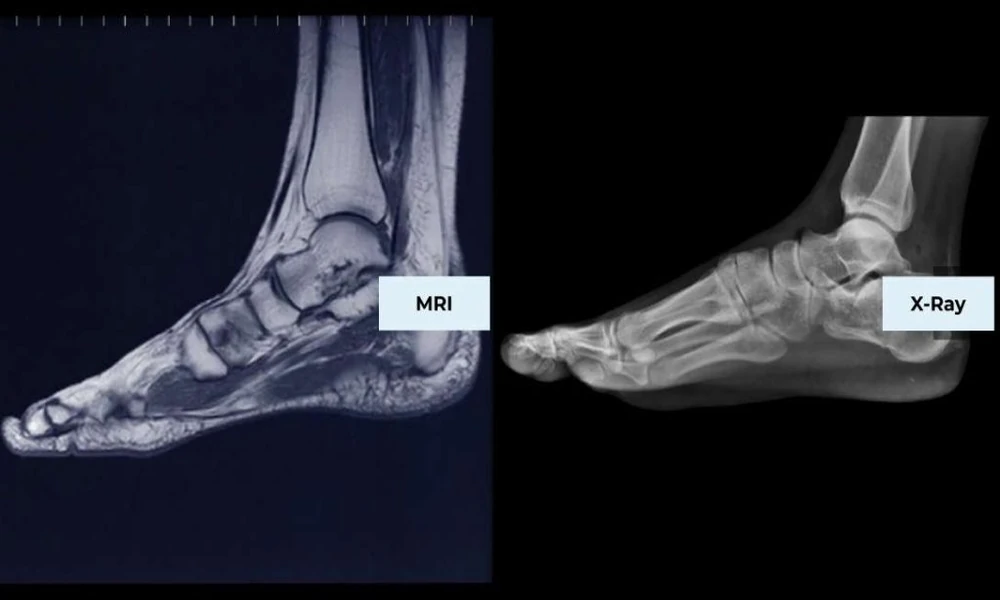




Foot and ankle injuries are among the most common problems in sports. Runners, basketball players, soccer players, and dancers place enormous repeated stress on these joints, and a single bad landing or a gradual buildup of training load can sideline an athlete for weeks.
Because so much depends on getting the diagnosis right, imaging often plays a central role in deciding how to treat an injury and when it is safe to return. MRI is one of the most useful tools for this, but it is not always the first step, and knowing when it helps can save time and frustration.
When an X-Ray Comes First
After an acute ankle or foot injury, X-rays are usually the starting point. They are quick, widely available, and very good at showing obvious fractures and joint alignment. Clinicians often use established guidelines, such as the Ottawa ankle rules, to decide whether an X-ray is needed after a sprain.
The limitation is that X-rays show bone far better than soft tissue. Ligaments, tendons, cartilage, and early bone stress do not appear clearly, so a normal X-ray does not always mean the injury is minor.
What MRI Shows That Other Imaging Can Miss
MRI produces detailed images of both soft tissue and bone without radiation. It can show ligament tears, tendon damage, cartilage injuries, fluid in the joint, and swelling inside the bone that signals a stress reaction or bruise.
For athletes with persistent pain after a seemingly simple injury, this detail often makes the difference between a vague diagnosis and a clear treatment plan. Clinicians requesting MRI of the foot and ankle usually want to answer a specific question about structures that X-rays cannot reveal.
Common Athletic Injuries Diagnosed With MRI
Ankle ligament injuries are the most familiar example. The lateral ligaments on the outside of the ankle tear in many rolled-ankle sprains, and MRI can grade the damage when an ankle stays unstable or painful longer than expected.
High ankle sprains, which involve the syndesmosis connecting the two lower leg bones, often take longer to heal than standard sprains. MRI helps confirm this injury, which matters because it can change the treatment plan and return-to-play timeline significantly.
Achilles tendon problems range from chronic tendinopathy to partial and complete tears. MRI shows the extent of tendon thickening, degeneration, or tearing, helping guide decisions about rest, rehabilitation, or surgery.
Peroneal tendon injuries on the outer ankle are sometimes missed and mistaken for lingering sprain pain. Persistent swelling and pain behind the outer ankle bone after a sprain is a common reason to look for them on MRI.
Stress fractures and bone stress injuries are frequent in runners and jumping athletes. Early on, X-rays are often normal, while MRI can detect bone marrow swelling well before a fracture line appears, allowing earlier treatment.
Cartilage injuries, particularly osteochondral lesions of the talus, can follow ankle sprains and cause deep, aching pain, catching, or swelling. Lisfranc injuries in the midfoot are another important diagnosis, since subtle ones can be overlooked and lead to long-term problems if untreated.
Why Timing Matters for Athletes
Athletes and coaches understandably want answers quickly. When symptoms do not improve as expected within a few weeks, or when an injury seems more severe than the initial exam suggested, an MRI can prevent a premature return to play that risks a worse injury.
At the same time, not every sprain needs an MRI. Many ankle injuries heal well with appropriate rest and rehabilitation, and imaging is most useful when it is likely to change treatment.
Weight-Bearing and Positional Imaging
The foot and ankle function under load, and some problems relate to alignment or instability that appears when standing. Most conventional MRI scans are taken with the foot at rest, which is fine for the majority of injuries.
For certain questions about alignment, arch collapse, or instability, some physicians consider weight-bearing imaging, which shows the joints while they support body weight. This is an evolving area, and the decision depends on what the clinician needs to see.
Preparing for a Foot or Ankle MRI
The exam is painless and usually takes about 30 to 45 minutes. Patients lie or sit still while the foot rests inside a coil designed to capture fine detail. Remove metal objects, and tell the staff about any surgical hardware, implants, or prior injuries involving metal fragments.
Most foot and ankle MRIs do not require contrast. In some cases, especially when evaluating cartilage or subtle joint problems, a physician may order MR arthrography, which involves injecting contrast into the joint before the scan.
Using Results to Guide Return to Play
An MRI result is most valuable when combined with a careful exam and an understanding of the athlete’s sport and goals. The same finding may lead to different decisions for a recreational runner and a collegiate sprinter.
Athletes should ask their clinician what the images show, how the injury is expected to heal, and what milestones need to be met before training resumes. Injuries often affect the whole leg as well, and pain in one joint can change how an athlete loads another, which is why some patients also need an MRI of the knee when compensation patterns develop.
Conclusion
MRI is one of the best tools for understanding foot and ankle injuries that X-rays cannot fully explain. It reveals ligament tears, tendon damage, bone stress, and cartilage injuries that shape treatment and recovery.
Used at the right time, it helps athletes avoid both unnecessary delay and a return to sport before the injury is ready, giving them the clearest path back to training and competition.